
Andrey Klymchenko receives an ERC grant to create structures similar to artificial neurons

Published on July 1, 2025

Following an ERC Consolidator Grant in 2015 and an ERC Proof of Concept Grant in 2020, Andrey Klymchenko has been awarded the ERC Advanced Grant. As a CNRS research director at the Bioimaging and Pathology Laboratory in Illkirch, the researcher will use this prestigious European funding to spend the next five years developing synthetic biomaterials capable of capturing and releasing molecules on command, much like artificial neurons. Although fundamental in nature, the research conducted by his laboratory opens up promising diagnostic and therapeutic prospects.

Nanochemistry and Bioimaging: Fluorescent Nanoscale Tools for Healthcare

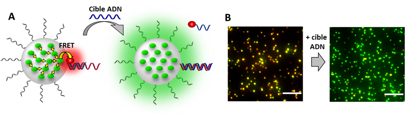
Since its founding in 2015, Dr. Andrey Klymchenko’s team has been developing and characterizing new molecular and supramolecular materials—fluorescent probes. These probes are tools capable of detecting and identifying molecules of interest within cells and biological fluids; their primary applications are imaging and diagnostics. For example, the team has developed nanoscale fluorescent probes that light up in the presence of DNA and RNA targets specific to certain diseases (Figure 1A,B)¹. In the future, these probes could be used to diagnose cancers and viral diseases by analyzing the fluorescence of the probes when they come into contact with patient samples. This technology led to the creation of the startup BrightSens Diagnostics in 2022, dedicated to developing simple and rapid medical tests that can be used directly with patients.

Figure 1: (A) Principle of the nanoparticle-based fluorescent probe for the detection of cancer cell-specific DNA biomarkers, and (B) its two-color response in the presence of the DNA target, as observed under a fluorescence microscope (each dot corresponds to a nanoprobe). 1

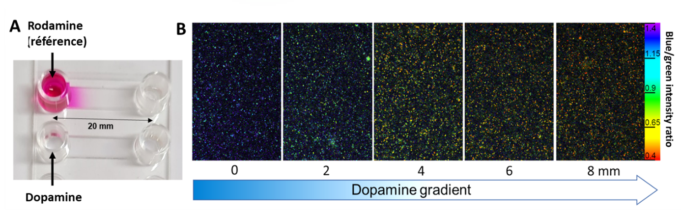
Recently, the team developed a novel chemical approach to quickly and effectively detect some of the smallest molecules in our bodies: neurotransmitters (such as dopamine)2. Artificial dopamine-specific receptors incorporate fluorescent molecules that change from red to blue in the presence of the neurotransmitter. This new, highly selective method makes it possible to quantify the amount of neurotransmitters present in the sample (Figure 2 A, B. For more details, readthe article dedicated to this technology).

Figure 2: (A) Creation of a neurotransmitter gradient in a microfluidic channel and imaging of this gradient using nanoparticle-based artificial receptors via fluorescence microscopy (B). The color gradually changes from red to blue depending on the dopamine concentration.2

Andrey Klymchenko now wants to take on a new challenge: transforming fluorescent probes into active materials capable of detecting and controlling, using light, the distribution of biologically active molecules. It is this ambitious project that has been selected by the European Research Council.

Precisely Controlling the Capture and Release of Molecules: A Bio-Inspired Material with Potential Medical Applications

Cells communicate with one another through signaling molecules, which bind to specific receptors on the surface of target cells. Our neurons also communicate in the same way by releasing neurotransmitters to receptors on the next neuron, thereby modulating its activity. Many diseases arise when the release and reuptake of these signaling molecules are disrupted. This is what happens, for example, in the brains of patients with depression, where certain neurotransmitters are released too quickly. A promising area of research in chemistry involves developing smart materials capable of replicating the behavior of neurons, thereby enabling chemical communication between materials and cells.

The CaptuRel project, funded by this new ERC grant, aims to develop biomaterials capable of capturing signaling molecules (such as neurotransmitters) and releasing them on command. The researcher explains the basics of this new type of material: “We will synthesize artificial receptors, also known as photocages, that can trap a target molecule when it passes within range (Figure 3), which is initially made possible by their accumulation within the biomaterial. This specific capture of neurotransmitters allows for their detection via fluorescence in a biological environment. To release the molecule, all that is needed is a light stimulus that induces a change in the receptor’s affinity. This CaptuRel biomaterial contains a multitude of photocages that capture natural molecules passing nearby in the biological environment. Once these molecules have accumulated, photorelease creates a chemical gradient near the material, which is necessary for communication with a neighboring cell.”

Figure 3: General Concept of the CaptuRel Project.

Although fundamental, this research could, in the long term, lead to medical devices capable of inducing controlled cellular responses: “Certain neurotransmitters play a key role in neuron growth and the formation of new neural connections. By precisely releasing this type of neurotransmitter, we will test whether this biomaterial can control neuron growth or guide connections between two neurons.” Once the experiments have been validated in vitro on isolated neurons, Andrey Klymchenko will continue the investigation on living tissue: “If all goes well, we plan to collaborate with neurobiology laboratories where CaptuRel could be tested locally on ex vivo brain tissue. That would already be a giant leap forward before we consider conducting tests on mice.”

With €2.5 million over five years, the researcher plans to hire four doctoral students and three postdoctoral researchers and acquire a state-of-the-art microscope needed for photomonitoring and imaging of biomaterials. Thanks to their long-term nature and substantial amount, these European grants allow scientists to spend less time seeking funding and more time on research itself. This is a welcome boost for the development of biomaterials with high therapeutic potential.

Sources

1. Melnychuk, N.; Klymchenko, A.S. DNA-Functionalized Dye-Loaded Polymeric Nanoparticles: Ultrabright FRET Platform for Amplified Detection of Nucleic Acids, J. Am. Chem. Soc. 2018, 140, 10856. DOI: 10.1021/jacs.8b05840


2. Kozibroda, B. Lehn, J.-M. Klymchenko, A. S. “Fluorescent Artificial Receptor for Dopamine Based on Molecular Recognition-Driven Dynamic Covalent Chemistry in a Lipid Nanoreactor,” Angew. Chem. Int. Ed. 2025, 64, e202419905. DOI: https://doi.org/10.1002/anie.202419905

Donation DONATIONS MAKE A DONATION