Highly Specific Artificial Dopamine Receptor: Toward the Medical Diagnosis of Brain Disorders

Published on February 24, 2025

In our bodies, changes in the concentration of certain molecules can serve as a warning sign of a developing disease, long before the first symptoms appear. The ability to accurately quantify the production of some of these molecules would make it possible to prevent a large number of diseases at very early stages.

The Photoactive Materials and Bioimaging team (University of Strasbourg – CNRS), led by Andrey Klymchenko in collaboration with Nobel laureate Jean-Marie Lehn, has developed a novel chemical approach to efficiently and rapidly detect some of the smallest molecules in our bodies: neurotransmitters. As chemical compounds released by neurons, neurotransmitters serve as indicators of our brain’s health. Measuring their levels in patients’ blood or urine makes it possible to detect neurodegenerative diseases (such as Parkinson’s or Alzheimer’s), depressive disorders, or epileptic syndromes.

A promising tool from both a medical and a basic research perspective

Andrey Klymchenko explains what makes this new method unique, as published in January 2025 in the journal *Angewandte Chemie International Edition*: “Currently, the methods used to measure neurotransmitters in patients’ urine are cumbersome; they must be performed in specialized laboratories and can be time-consuming. We have developed a concept for an artificial receptor that could, in the future, make it possible to analyze a large number of neurotransmitters at the same time, in just a few minutes.”

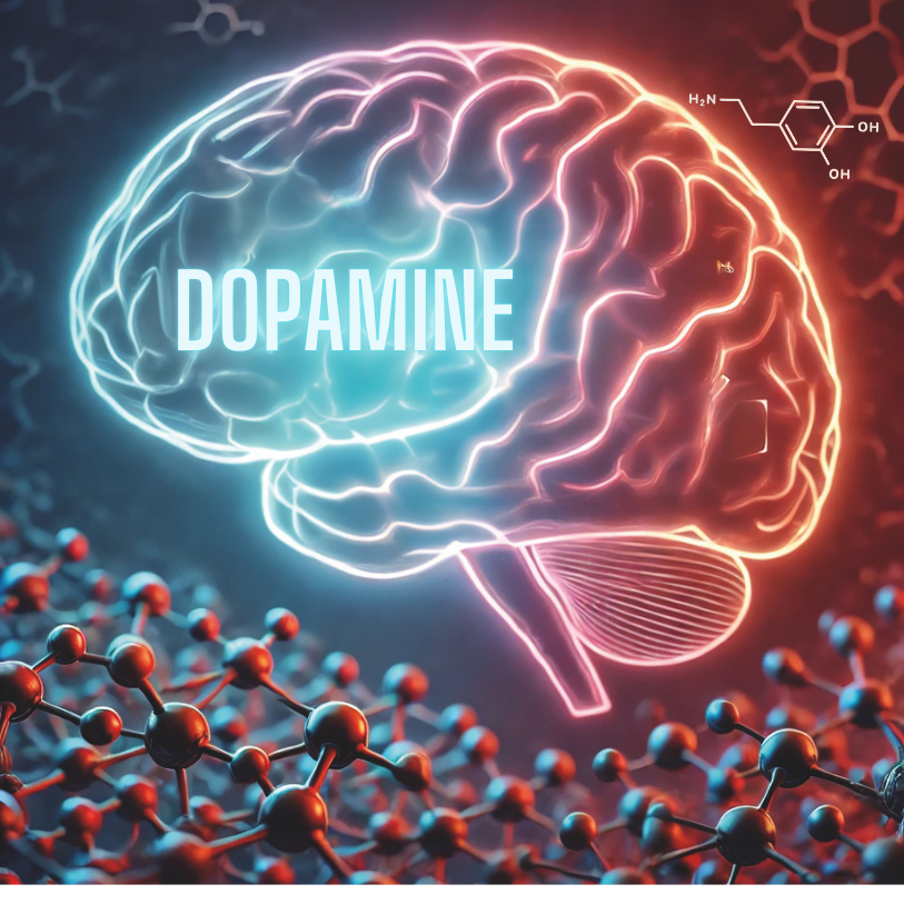
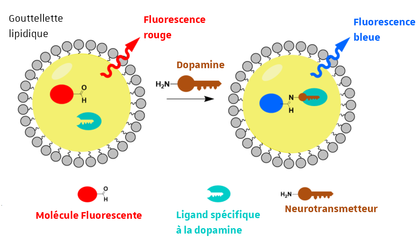
The team, which specializes in the design of fluorescent molecules and nanomaterials, has developed an ingenious supramolecular system for measuring dopamine—a neurotransmitter involved in numerous diseases—using fluorescence : “We designed the nanoscale artificial receptor. It consists of a fluorescent molecule that emits red light, but once bound to a chemical group characteristic of neurotransmitters (in this case, dopamine), its structure changes and the molecule emits blue light. This technique allows us to visualize and quantify dopamine concentrations, but it wasn’t specific—we had to devise a system so that only dopamine would be recognized (and not other neurotransmitters). To do this, we placed these fluorescent molecules inside nanoscale lipid droplets; no neurotransmitter can penetrate them without a little help. This is where a molecule (ligand) comes into play; it recognizes dopamine within the droplets with high specificity (like a key in a lock) and facilitates its entry so that it can bind to the fluorescent molecule.”

The Principle of the Artificial Receptor. 

In the absence of dopamine, the fluorescent molecule emits red light. In the presence of the neurotransmitter, the recognition molecule binds to the dopamine and allows it to be incorporated into the droplet. The dopamine can then bind to the fluorescent molecule, which then emits blue light.

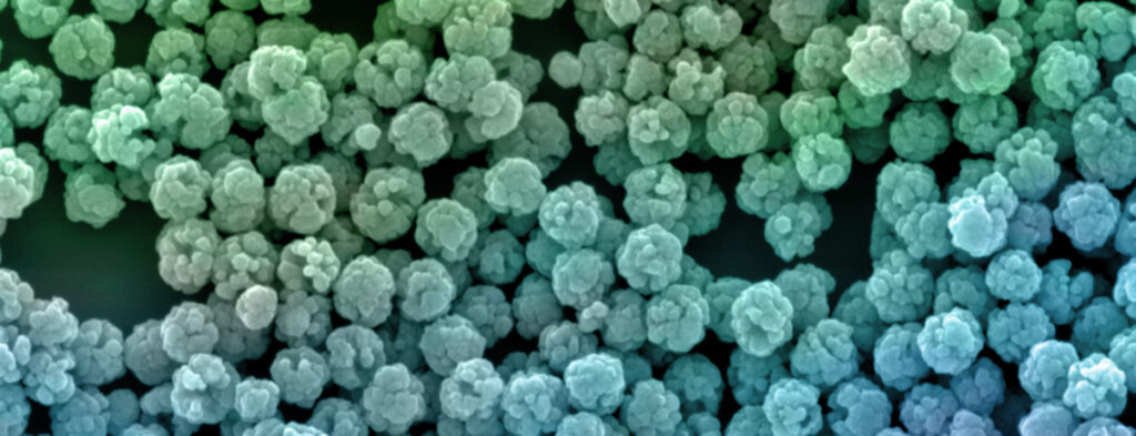
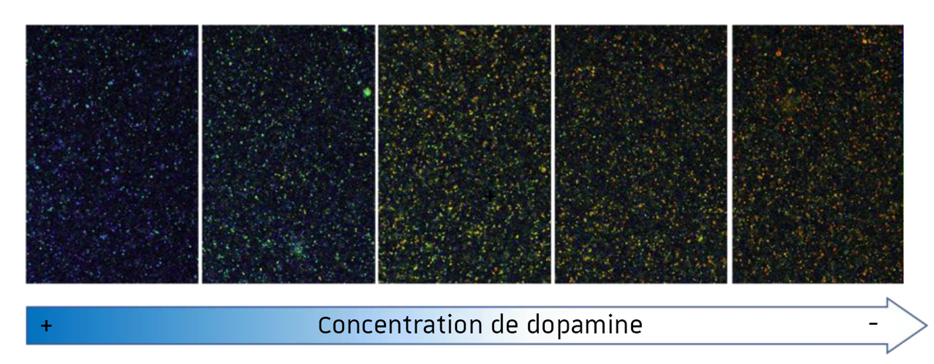
Microscopic images of nanodroplets as a function of dopamine concentration. The blue-to-red ratio varies as expected: blue droplets at high dopamine concentrations on the left, and a large number of red droplets at low concentrations on the right (the intermediate colors reflect a mixture of red and blue).

The researcher explains that the experiment was conducted in the presence of numerous other neurotransmitters to test the specificity of the method: “Only dopamine penetrates the droplets; this is a highly specific detection method.” We now have a valid proof of concept demonstrating the effectiveness of this new tool. The advantage of this method is that we will soon be able to test whether it works for other neurotransmitters by designing a specific receptor for each one.”


More than just a diagnostic tool, this new technique could also be developed to visualize the specific production of each neurotransmitter directly in living organisms. The goal would be to better understand neuronal activity in animal models without having to create genetically modified organisms. Indeed, these nanodroplets could be injected near the neurons under study to measure their activity using fluorescence microscopy to visualize the released neurotransmitters.

A High-Risk Project: A Bold Move Pays Off

Developing this technique, which had never been attempted before, was clearly a risky venture . “It’s difficult to secure funding to develop a technique like this; we had no guarantee that it would work.” Together with Prof. Jean-Marie Lehn, we were able to co-fund the thesis of the student, Bohdan Kozibroda, who conducted the experiments—a brilliant student from the University School of Research in Chemistry (SysChem), which the Foundation supports. Last year, my team also received substantial funding from the Foundation to develop this new concept. At every stage of this project, the Foundation was there to support us in one way or another.” As the recipient of a grant from the Jean-Marie Lehn Foundation, Andrey Klymchenko plans to explore the possibility of applying his method to other neurotransmitters. These funds will also enable him to improve the affinity of his receptor in order to detect dopamine at lower concentrations, closer to physiological levels.

In the medium term, the researcher makes no secret of his ambition to bring this project to fruition. This isn’t the researcher’s first attempt; he has already founded two startups: BrightSens Diagnostics, which designs fluorescent nanoparticles to detect cancers and viruses, and AstraNICE, which is developing a fluorescent coating to help surgeons better visualize organs during surgeries.

Source

Fluorescent Artificial Dopamine Receptor Based on Molecular Recognition-Driven Dynamic Covalent Chemistry in a Lipid Nanoreactor. Kozibroda, B., Lehn, M., & Klymchenko, A. S. Angewandte Chemie International Edition, January 13, 2025.

DOI: https://doi.org/10.1002/anie.202419905

Donation DONATIONS MAKE A DONATION